Funduscopy
Mydriasis is about the dilation of the pupil. Normally, the size of the pupil changes due to brightness or darkness.The pupil becomes small (Miosis) in bright places, and large in the dark.
This test is examined while the pupil is large by dilating drops (Mydriatic).
When it is necessary for the doctor to check the condition of the patient's lenses, this test is performed.

Diseases that require Mydriasis
Fundus diseases require Mydriasis.
This is because the test makes it able to examine the edge of the fundus.
Typical diseases that require Mydriasis are retinal detachment, cataract, macular hole, diabetic maculopathy, age-related macular degeneration, and macular edema.
This is because the test makes it able to examine the edge of the fundus.
Typical diseases that require Mydriasis are retinal detachment, cataract, macular hole, diabetic maculopathy, age-related macular degeneration, and macular edema.
After Mydriasis
In this test, the doctor examines the optic nerve and the retina by an ophthalmoscope.
This examination is able to check whether you have retinal disease.
This examination is able to check whether you have retinal disease.
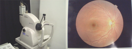
Fundus taken by the Fundus camera
This test takes about 5 minutes.


OCT

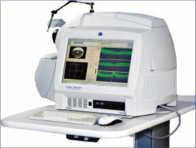
The OCT (Optical Coherence Tomography) is an instrument, which examines the tomographic image of the retina.
The result of this examination shows the condition of the retina in detail, and helps to decide treatment plans.
This test takes about 5-10 minutes.


