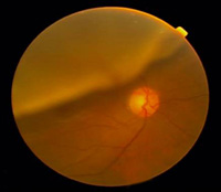
Retinal detachment describes an emergency in which a thin layer of tissue (the retina) at the back of the eye pulls away from its normal position.
Cause
Aging
As you age, the gel-like material that fills the inside of your eye, known as the vitreous, may change in consistency and shrink or become more liquid.
Previous severe eye injury
Diabetes
The prolonged levels of high blood sugar damage the small blood vessels within the retina. This may cause hemorrhages, exudates and even swelling of the retina.
Symptoms
Retinal detachment itself is painless. But warning signs almost always appear before it occurs or has advanced, such as:
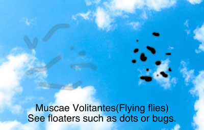
・The sudden appearance of many floaters — tiny specks that seem to drift through your field of vision
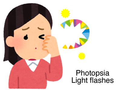
・Flashes of light in one or both eyes
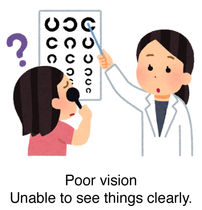
・Blurred vision
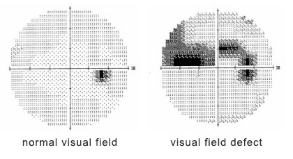
・Gradually reduced side (peripheral) vision
・A curtain-like shadow over your visual field
Testing
Funduscopic Examination
This is an examination to check the condition of your retina. We put eye drops that dilate your pupils and observe the retina and optic nerve to check if there aren’t any diseases.
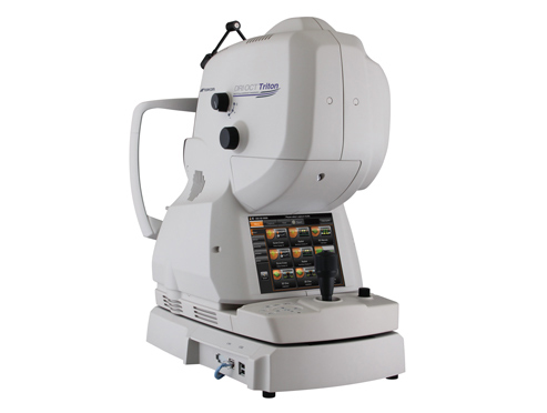
Optical coherence tomography (OCT)
It also helps your doctor look at the retina. A machine scans the back of the eye and provides detailed three-dimensional pictures of the retina. This helps measure retinal thickness and find swelling of the retina.
Treatment
Laser coagulation
Laser coagulation uses the heat from a laser to seal or destroy abnormal, leaking blood vessels in the retina. Laser coagulation may be used to prevent further progression of retinopathy.
Surgery
If your retina has detached, you'll need surgery to repair it, preferably within days of a diagnosis. The type of surgery your surgeon recommends will depend on several factors, including how severe the detachment is.
After surgery your vision may take several months to improve. Some people never recover all their lost vision.
We will write a referral letter and refer you to another hospital, if you need.


