The retina is the inner lining of the eye; it is the thin, light-sensitive tissue that generates vision. Tears can form in the retina, creating a risk of retinal detachment such as Atrophic hole and Tractional hiatus.
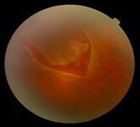
Cause
Aging
The vitreous is a clear gel-like substance that fills in the back cavity of the eye, which is lined by the retina. At birth, this gel is attached to the retina, but as we age, the gel separates from the retina creating a posterior vitreous detachment or PVD.
High myopia
People with high myopia have longer eyes (axial elongation), which means that the retina is a more stretched and therefore prone to peripheral retinal hole.
Eyeball bruise
Symptoms
Retinal detachment itself is painless. But warning signs almost always appear before it occurs or has advanced, such as:
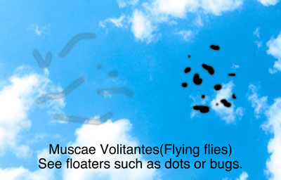
・The sudden appearance of many floaters — tiny specks that seem to drift through your field of vision
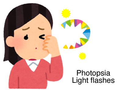
・Flashes of light in one or both eyes
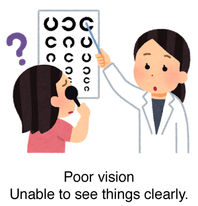
・Blurred vision
・Gradually reduced side (peripheral) vision
・A curtain-like shadow over your visual field
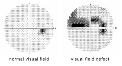
Testing
Funduscopic Examination
This is an examination to check the condition of your retina. We put eye drops that dilate your pupils and observe the retina and optic nerve to check if there aren’t any diseases.
Treatment
Laser coagulation
Laser coagulation uses the heat from a laser to seal or destroy abnormal, leaking blood vessels in the retina. Laser coagulation may be used to prevent further progression of retinopathy.
Please feel free to visit our clinic.
(If you take reservation to visit our clinic, we give priority to your examination order.)


